- Home
- Research
- Achieving Excellence - Research at Washington University Orthopedics
- Understanding Low Back Pain
Understanding Low Back Pain
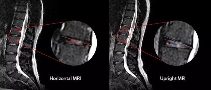
An upright MRI (right) versus a traditional horizontal MRI (left) can more clearly identify differences in the spinal structure, helping clinicians to further understand the reason for low back pain.
Can standing upright while getting an MRI really make a difference in the diagnosis and treatment of low back pain? At Washington University School of Medicine, one orthopedic researcher believes the answer could be yes!
Simon Tang, PhD, has been researching what he calls a “head-scratcher of a group” – young adults between the ages of 18 and 30 who have physically healthy backs but who experience bouts of low back pain.
“These are patients who do not have degenerative disc disease or spondylosis (osteoarthritis of the spine), but they develop low back pain symptoms when they stand for long periods of time,” he says. “When they move or lean on something, the pain goes away. But one thing is consistent. Over time, their symptoms can lead to chronic low back pain three to five years later. By understanding how this group develops low back pain, we can better understand how low back pain initiates, which then could give clinicians a unique opportunity to intervene early.”
Tang’s lab is focused on identifying disease mechanisms that impact the structure-function relationships of skeletal tissues. He is both a bioengineer and an orthopedic researcher. The combined expertise makes him ideal to examine the forces that impact the skeletal system.
Tang and his co-investigator Linda Van Dillen, PT, PhD, wanted to know why low back pain occurred in this healthy young adult group only when they were standing. In collaboration with the Open Upright MRI of Missouri in Creve Coeur, they began imaging healthy patients to see if there are significant changes in their anatomy and soft tissues using a magnetic resonance imaging (MRI). Using upright magnetic resonance imaging (MRI), they began imaging healthy patients to see if there were significant changes in their anatomy or soft tissue. Patients were asked to stand for up to two hours in an upright MRI so that researchers could monitor the spinal changes as low back pain occurred. Those images were then compared to initial standing MR images when there is no pain.
“We noticed that the spinal alignment and curvature were different in the standing images than in those taken while patients were lying down,” says Tang. “We know the back is relaxed when lying down but we were surprised to see the local structural differences that we could actually quantify in the upright MR images.”
What they found was that spinal curvature increased when patients stood up. More significantly, they also found that the increased curvature was not always distributed evenly over the entire spine, meaning that there were small changes in some spinal segments but big changes in other segments. “This could pre-dispose someone to back pain,” explains Tang. “These segments would experience greater changes in loading and fatigue.”
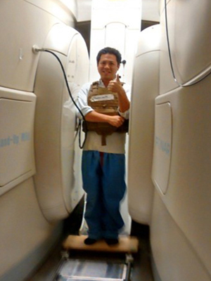
Simon Tang, PhD, demonstrating an open upright MRI.
Their preliminary data seems to suggest that gravity and anatomy all play significant roles in low back pain in young adults. It also points to the use of upright MRI as a way to identify optimal treatments for low back pain.
“We hope that these findings can inform clinical decisions by identifying the spine segments that may be at higher risk of pain and guide potential interventions before symptoms worsen. One strategy could be to engage in postural retraining or strengthening exercises that target those spine segments,” Tang says.
Physicians also could utilize these MR images for computer modeling prior to any surgical correction to identify a loading situation that is optimal for each patient. “Even very small angular changes of one to two degrees in the intervertebral disc angles can make a significant difference in the stresses experienced by those discs,” he notes.
Tang and Van Dillen are recruiting more patients into their study. Within the next several years, Tang says, “I think these findings could be translated to the clinical setting. It would be very easy for doctors to prescribe a standing MRI if it will help them make better decisions and result in better patient outcomes.” This study is funded by the National Institutes of Health.
Next article: Young Adult Hip Deformities